What is thoracic outlet syndrome?
The thoracic outlet is the space between the first rib and the collar bone. There are muscles, nerves and blood vessels that pass through this space. Thoracic outlet syndrome is where nerves and/or blood vessels in this space become squashed. Typically, this compression happens when your arm is in certain positions. This can cause symptoms in your arm such as swelling, pins and needles, pain, or a change in colour.
What is an ultrasound scan of the arteries in your arm?
An ultrasound scan of your arteries may also be called an 'arterial duplex' or 'arterial Doppler' of your arms. This test uses ultrasound to produce images of the arteries in your arms. The arteries carry blood away from the heart to supply the extremities.
It is a safe and effective way to assess the blood flow and can determine whether there are any narrowings or blockages in the arteries. It can also determine whether the blood vessel is being compressed within the thoracic outlet space. This will allow the consultant to appropriately plan your treatment for you.
The test is painless and does not use any radiation or needles. There are no risks associated with it.

About the test
Where do I go?
The Vascular Studies Unit (VSU) is on level 5 of the Addenbrooke's Treatment Centre (ATC). Please inform reception of your arrival straight away. There are often other clinics in progress so you may not be called in order of arrival.
What is consent?
Before your test is performed you must give your consent or permission. Consent is the process by which you give permission to healthcare professionals to provide your care and treatment. It may be implied (offering your arm for a blood pressure reading) or formal (signing a formal consent form for an operation). In either case your consent must be given voluntarily, and you must have all the information you need to make a decision. If you feel you do not have enough information or do not understand the procedure, please ask.
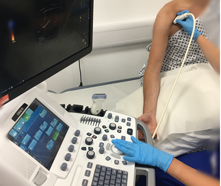
How is an ultrasound scan of the arteries in your arm performed?
There is no preparation required and you may eat and drink as usual prior to the test. A clinical vascular scientist (who might be male or female) will perform and interpret your ultrasound scan. The scan will be performed from the wrist to the neck, so you may be asked to remove your top so the scientist can scan the area of interest. The lights will be dimmed to allow the best images to be obtained.
The scan will be performed with you lying down or seated on the edge of the couch. Gel is applied to your arm and the ultrasound probe will be moved along the arm to view all of the arteries.
You will be asked to put your arm in various positions during the scan. The scan takes approximately 30 to 60 minutes. During the test, you may hear some 'swooshing' noises from the ultrasound machine. These sounds are normal.
What happens next?
The clinical vascular scientist can comment briefly on the findings and will write a report for the consultant who requested the test. You will be able to discuss the results of this investigation fully with the referring team at your next outpatient appointment. In rare cases, the clinical vascular scientist may need to discuss the result with a doctor before you leave.
Other important information
You may bring a relative or friend in with you during the test or request a chaperone if you would like one.
If you require further information, please do not hesitate to contact the Vascular Studies Unit (VSU) on 01223 348117.
Privacy and dignity
You may bring a relative or friend in with you during the test or request a chaperone if you would like one.
Same sex bays and bathrooms are offered in all wards except critical care and theatre recovery areas where the use of high-tech equipment and / or specialist one-to-one care is required.
We are smoke-free
Smoking is not allowed anywhere on the hospital campus. For advice and support in quitting, contact your GP or the free NHS stop smoking helpline on 0800 169 0 169.
Other formats
Help accessing this information in other formats is available. To find out more about the services we provide, please visit our patient information help page (see link below) or telephone 01223 256998. www.cuh.nhs.uk/contact-us/accessible-information/
Contact us
Cambridge University Hospitals
NHS Foundation Trust
Hills Road, Cambridge
CB2 0QQ
Telephone +44 (0)1223 245151
https://www.cuh.nhs.uk/contact-us/contact-enquiries/