A team effort to develop the world’s first cot-side functional neuroimaging technology to speedily detect brain injuries including cerebral palsy in newborn babies is being spearheaded in Cambridge.
The three-year Fast UltraSound Imaging with Optics in the Newborn (fUSiON) study, involving high-risk infants, is due start at Cambridge University Hospitals NHS Foundation Trust (CUH). If the study is successful, the wearable device, which fits like a swimming cap, could become a common in every UK hospital.
Brain injury in the newborn is a major cause of lifelong disability. This includes conditions such as cerebral palsy, learning difficulties, epilepsy, and autism spectrum disorders. These result in significant physical, psychological, and economic burden on individuals, their families, and society.
Structural brain imaging with cranial ultrasound (CUS) and magnetic resonance imaging (MRI) remain the traditional way of imaging the brain in sick and preterm infants. However, both techniques are limited in their ability to predict the extent or nature of injury and any future impairment. This is because there is a complex relationship between brain structure and brain function. Furthermore, MRI scans are costly and cumbersome, meaning they cannot be performed at regular intervals.
Watch: Fusion Study
Link: https://youtu.be/Qnk0-1tcYsM
Therefore, the ability to measure whole brain function at the cot-side will represent a major step forward in the ability of clinicians to identify those infants at risk of problems in a timely manner. This will potentially enable more targeted therapies to be started at an early stage for optimising neurodevelopmental outcomes.
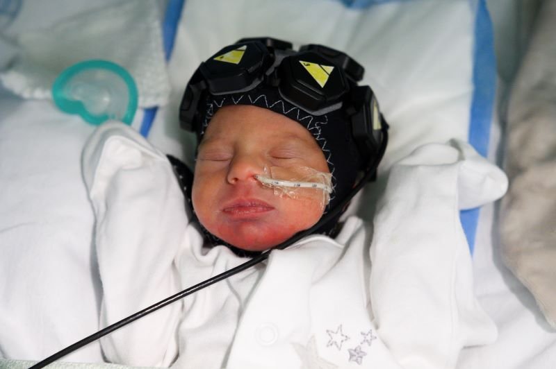
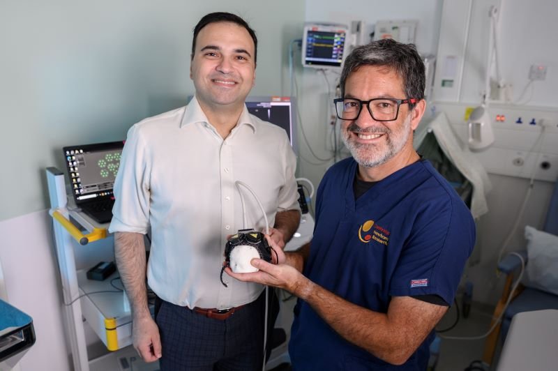
The specialised cap is fitted with multiple light sensors that detect changes in blood oxygenation on the surface of the brain – a technology known as high-density diffuse optical tomography – or HD-DOT. It is similar to the widely-used devices put on a baby’s toe post-birth to monitor oxygen levels.
The cap also includes a special kind of ultrasound, called functional ultrasound (fUS), which captures data at a very high frequency for enabling images of small blood vessels (microvasculature) deep in the brain to be generated.
Each technique is good at detecting activity in different parts of the brain: HD-DOT works well in superficial areas, whilst fUS is good for visualising deeper structures. The project aims for the first time to combine both technologies to obtain whole brain activity measurements at the cot-side.
The study, all done with parents’ consent, is being led by a team at Cambridge University Hospital’s neoLAB, which is based in the Evelyn Perinatal Imaging Centre at the Rosie Maternity Hospital, which sits alongside Addenbrooke’s Hospital.
The work is partly funded by a prestigious Marie Skłodowska-Curie Postdoctoral European Fellowship under the Horizon Europe Framework Programme for Research and Innovation awarded to Dr Flora Faure in the Department of Clinical Neurosciences, University of Cambridge.
The NIHR HealthTech Research Centre (HRC) in Brain Injury is also supporting the project by funding an additional PhD studentship, as well as wider technical, clinical, and implementation expertise.
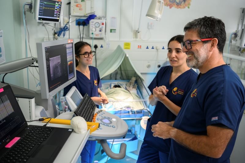
Professor Topun Austin is a consultant neonatologist and director of the Evelyn Perinatal Imaging Centre. He is also the Life Course Theme Lead for the Brain Injury HRC, exploring brain treatments at the extremes of life – young and old.
He explained:
The fUSiON Study aims to develop and demonstrate a system for the cot-side assessment of brain activity in newborn infants and is currently the first of its kind in the world.
We have spent 12 months successfully proving the concept with the help of healthy and premature babies, and will now move on to the second phase, which focuses on those babies considered to be at higher risk of brain damage.
Understanding and looking at brain activity patterns in both term and preterm infants can help us identify infants most vulnerable to injury at an early stage.
Professor Topun Austin
The Brain Injury HRC Co-Director, consultant academic neurosurgeon, Dr Alexis Joannides, said:
We are delighted to support this innovative work, bringing together the excellent track record of the neoLAB team with the expertise and cutting-edge technologies of our collaborators in London and Paris.
The neonatal cap has potential to transform the way we diagnose, monitor, and understand brain injury in newborn babies. If successful, it could offer a cost-effective imaging tool for routine use in hospitals across the UK and beyond.
Dr Alexis Joannides
The Cambridge team is working in partnership with the DOT-HUB research group at University College London (UCL) and and Gowerlabs, developers of the LUMO wearable HD-DOT system.
Experts from the Physics for Medicine group based at the Ecole Supérieure de Physique et de Chimie Industrielles (ESPCI), Paris, are pioneers in the field of functional ultrasound.
Dr Samuel Powell, CTO of Gowerlabs, said:
Our previous collaborations with neoLAB have demonstrated the feasibility of HD-DOT to study the newborn brain. It is exciting to be continuing this collaboration in combining two complementary technologies with potential real world benefits for sick and premature babies.
Dr Samuel Powell
Dr Charlie Demené from the Physics for Medicine group in Paris, added:
We are pleased to contribute our considerable expertise and experience and to be part of this international collaboration
Dr Charlie Demené
Anyone who wants to be part of the fUSiON study’s new Patient Advisory Panel, or is a parent who thinks their baby may benefit once born, should email info@brainhrc.org.
The study comes as work continues to build Cambridge Children’s Hospital, which will be unique in treating mental and physical health together under one roof, alongside research. To make a donation, visit Addenbrooke’s Charitable Trust (ACT).